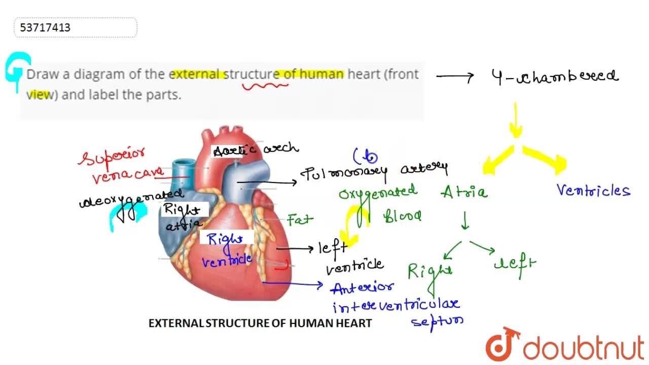
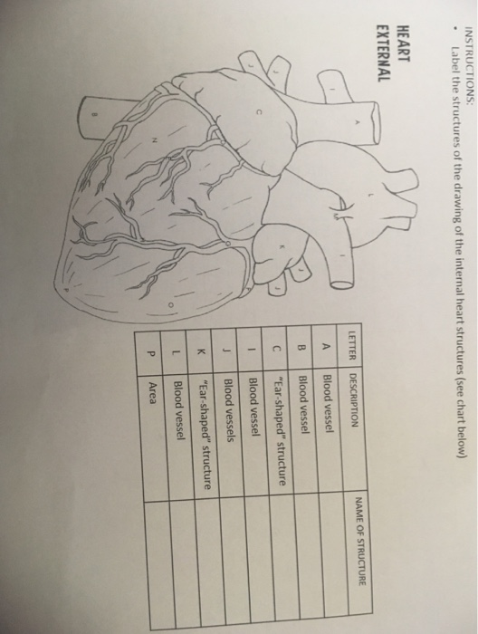
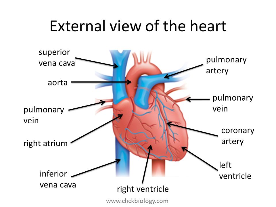
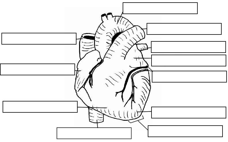
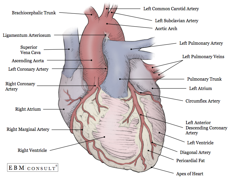
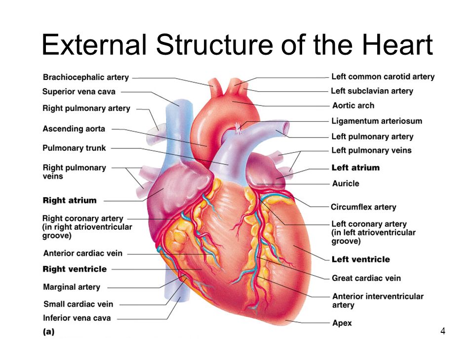
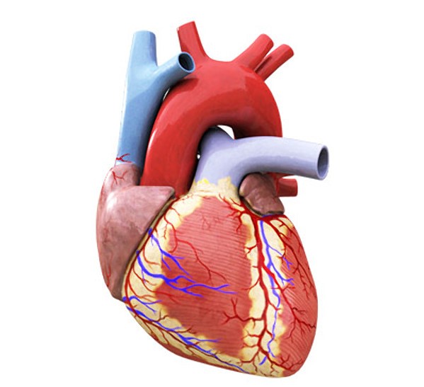
41 external structure of the heart with labels
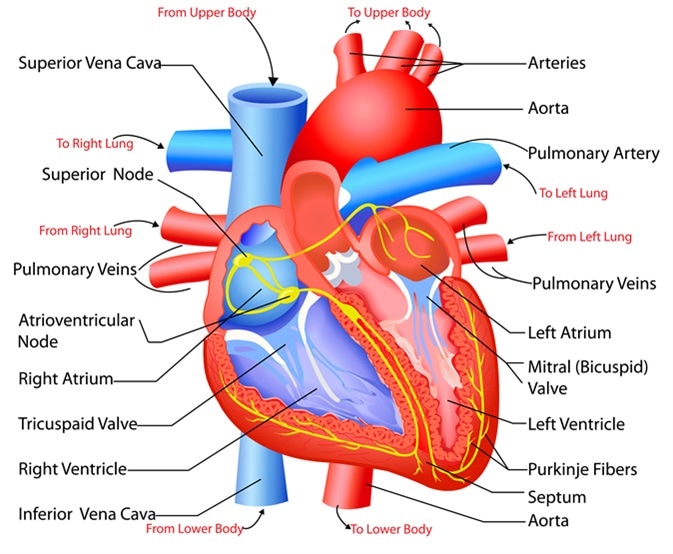
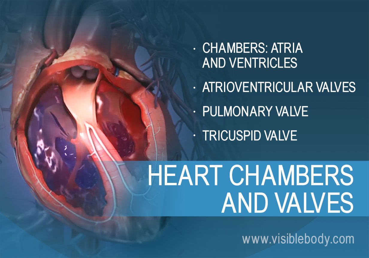
The Location, Size, and Shape of the Heart | GetBodySmart The heart is located underneath the sternum in a thoracic compartment called the mediastinum, which occupies the space between the lungs. The sternum and mediastinum. 1 2 3 Previous Next It is approximately the size of a man's fist (230-350 grams) and is shaped like an inverted cone. heart | Structure, Function, Diagram, Anatomy, & Facts A thin layer of tissue, the pericardium, covers the outside, and another layer, the endocardium, lines the inside. The heart cavity is divided down the middle into a right and a left heart, which in turn are subdivided into two chambers. The upper chamber is called an atrium (or auricle), and the lower chamber is called a ventricle.
Structure and Function of the Heart - News-Medical.net The heart wall is composed of three layers, including the outer epicardium (thin layer), middle myocardium (thick layer), and innermost endocardium (thin layer). The myocardium is made up of...

External structure of the heart with labels
Frog Anatomy & Physiology: Learn About All Parts Of The Frog The circulatory system of the frog consists of a three-chambered heart, blood, blood vessels, and the spleen. The frog's heart has two upper chambers (atria) and one lower chamber known as the ventricle. The right atrium receives oxygen-poor blood from the body, and the left receives oxygen-laden blood from the lungs. How the Heart Works - The Heart | NHLBI, NIH - National Institutes of ... The Heart. The heart is an organ about the size of your fist that pumps blood through your body. It is made up of multiple layers of tissue. Your heart is at the center of your circulatory system. This system is a network of blood vessels, such as arteries, veins, and capillaries, that carries blood to and from all areas of your body. Cat Anatomy and Physiology 101: All You Need to Know A - cervical bones, B - thoracic bones, C - lumbar bones, D - sacral bones, E - tail bones, 1 - cranium, 2 - mandible, 3 - scapula, 4 - sternum, 5 - humerus, 6 - radius, 7 - phalangeals, 8 - metacarpals, 9 - carpal bones , 10 - ulna, 11 - ribs, 12 - patella, 13 - tibia, 14 - metatarsals, 15 - tarsal bones, 16 - fibula, 17 - femur
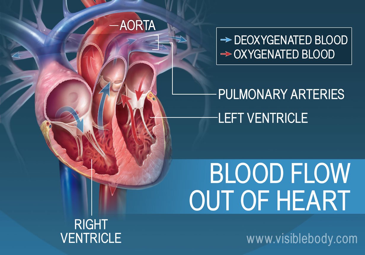
External structure of the heart with labels. Heart - Wikipedia The heart has four chambers, two upper atria, the receiving chambers, and two lower ventricles, the discharging chambers. The atria open into the ventricles via the atrioventricular valves, present in the atrioventricular septum. This distinction is visible also on the surface of the heart as the coronary sulcus. [18] Heart anatomy: Structure, valves, coronary vessels | Kenhub The heart is shaped as a quadrangular pyramid, and orientated as if the pyramid has fallen onto one of its sides so that its base faces the posterior thoracic wall, and its apex is pointed toward the anterior thoracic wall. Heart: Anatomy | Concise Medical Knowledge - Lecturio The heart is a 4-chambered muscular pump made primarily of cardiac muscle tissue. The heart is divided into 4 chambers: 2 upper chambers for receiving blood from the great vessels, known as the right and left atria, and 2 stronger lower chambers, known as the right and left ventricles, which pump blood throughout the body. Skin Layers: Structure, Function, Anatomy, and More - Verywell Health The stratum lucidum is a separate layer only in the thicker epidermis on the palms of the hands and soles of the feet. In thinner areas, its cells and functions are incorporated into other layers. The stratum lucidum: Allows the skin to stretch Contains a protein that helps skin cells degenerate
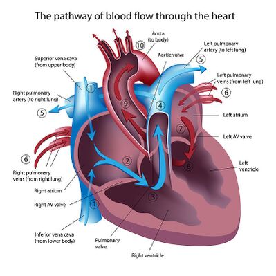
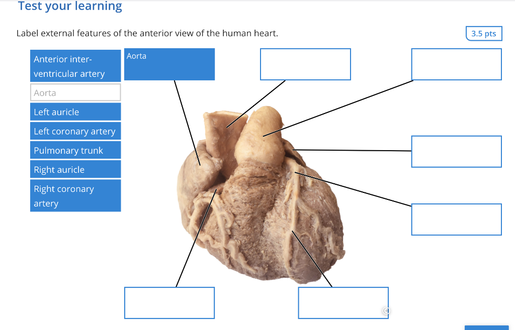
Histology, Heart - StatPearls - NCBI Bookshelf The heart is a four-chambered organ responsible for pumping throughout the body. It receives deoxygenated blood from the body, sends it to the lung, receives oxygenated blood from the lungs, and then distributes the oxygenated blood throughout the body. At the histological level, the cellular features of the heart play a vital role in the normal function and adaptations of the heart. Correctly Label The Following External Anatomy Of The Anterior Heart ... The external anatomy of the human heart consists of the four chambers that form the apex of the heart. Each chamber has an apex that corresponds to a box. There are two boxes on each side of the heart: the atria and the ventricles. The left atrium is a branching organ. The pulmonary trunk contains the aorta and pulmonary veins. Labeled imaging anatomy cases | Radiology Reference Article ... This article lists a series of labeled imaging anatomy cases by body region and modality. Brain CT head: non-contrast axial CT head: non-contrast coronal CT head: non-contrast sagittal CT head: angiogram axial CT head: angiogram coronal CT... The Ultimate Heart Model & Sheep Heart Practice Quiz! As is the case in the lab practical, each correct answer counts. So, make sure you learn from the feedback. Questions and Answers 1. 1. Name the structure- be specific. 2. 2. Name the vessel- be specific. 3. 3. Name the structure-be specific. 4. 4. Name the vessels. 5. 5. Do the vessels in #4 carry blood to or from the heart? A. To the heart B.
Heart: illustrated anatomy - e-Anatomy - IMAIOS This interactive atlas of human heart anatomy is based on medical illustrations and cadaver photography. The user can show or hide the anatomical labels which provide a useful tool to create illustrations perfectly adapted for teaching. Anatomy of the heart: anatomical illustrations and structures, 3D model and photographs of dissection. Heart Labeling Quiz: How Much You Know About Heart Labeling? Here is a Heart labeling quiz for you. The human heart is a vital organ for every human. The more healthy your heart is, the longer the chances you have of surviving, so you better take care of it. Take the following quiz to know how much you know about your heart. Questions and Answers 1. What is #1? 2. What is #2? 3. What is #3? 4. What is #4? Learn the Arteries and Veins of the Circulatory System - The Biology Corner This unit takes approximately three days: Arteries and veins of the heart, neck, and arms. Arteries branching from the abdominal aorta. Veins branching from the inferior vena cava. Finally, a day can be reserved for review and labeling practice. I have anatomy coloring books that I sometimes copy relevant pages for students to color. Lab 2: Anatomy of the Heart - Anatomy & Physiology: BIO 161 / 162 ... Lab 2: Anatomy of the Heart. A&P Lab Manual. Lab Atlas: Heart. Lab 2: Anatomy of the Heart. Additional Activities: Lab 2. Models of the Heart - Blank. Models of the Heart - Labeling Activity. Practice Quiz. Heart Anatomy Practice Quiz . Lab Model Videos. Sheep Heart Dissection. Plastic Heart Model Anatomy.
Heart Wall Anatomy | Structure of the Heart Wall | GetBodySmart The epicardium is a serous membrane that consists of an external layer of simple squamous and an inner layer of areolar tissue ( loose connective tissue ). The squamous cells secrete lubricating fluids into the pericardial cavity. The thick middle layer of the heart wall is called the myocardium.
What Are the 5 Parts of the Integumentary System? - MedicineNet Center. The 5 parts of the integumentary system—skin, hair, nails, glands, and nerves—protect the body from environmental elements. The integumentary system is made up of organs and structures that protect the inside of the body from environmental elements. The 5 parts of the integumentary system include:
Anatomy, Arterioles - StatPearls - NCBI Bookshelf Blood circulates through the body via the vascular tree consists of arteries, veins, and capillary beds. An artery carries blood away from the heart, and distribute throughout the body by its succeeding smaller branches. Eventually, the smallest branch of the artery is called arterioles, which further divide into tiny vessels to form the capillary bed. Nutrients and wastes exchange between the ...
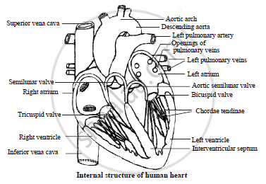
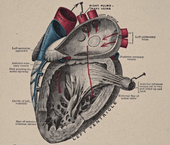
Internal Anatomy Of The Heart - Heart Failure - GUWS Medical A cross section cut through the heart reveals three layers (Fig. 7): (1) a superficial visceral pericardium or epicardium (epi = "upon" + "heart"); (2) a middle myocardium (myo = "muscle" + "heart"); and (3) a deep lining called the "endocardium" (endo = "within," derived from the endoderm layer of the embryonic trilamina).The endocardium is a sheet of epithelium called endothelium that rests ...
Human heart: Anatomy, function & facts | Live Science The human heart is an organ that pumps blood throughout the body via the vessels of the circulatory system, supplying oxygen and nutrients to the tissues and removing carbon dioxide and other ...
Surface projections of the heart: Borders and landmarks | Kenhub The right margin of the heart consists of two lateral convex arches separated by an obtuse angle. In adults, the lower arch belongs to the right atrium, while the upper arch represents the ascending aorta. However, in children, the upper arch is actually flat and is formed by the superior vena cava.
Kidney Structures and Functions Explained (with Picture and Video) The glomerulus connects to a long, convoluted renal tubule which is divided into three functional parts. These consist of the loop of Henle (nephritic loop), the proximal convoluted tubule, and the distal convoluted tubule, which empties into the collecting ducts. These collecting ducts fuse together and enter the papillae of the renal medulla.
External Structures of Animals: Lesson for Kids - Study.com All animals have external structures, which means outside parts of the body. Most animals have a head, body covering, limbs, and some form of a tail. Although these body parts may look different on...
Clavicle (Collarbone) - Location, Anatomy, & Labeled Diagram Along with the rib cage, they protect the heart from external shock. Anatomy of the Parts of Clavicle With its Bony Landmarks. Being a long bone, it has two ends, the sternal and acromial ends. The region in between the two ends is known as the shaft. C lavicl e. 1. Sternal (Medial) End.
The Ear: Anatomy, Function, and Treatment - Verywell Health External acoustic meatus: This is the bone and cartilage lined canal that leads from the outside to the inside of the ear. Its outer portion is surrounded by cartilage, and the inner part is surrounded by bones of the skull. This portion curves, slightly up and to the back, before bending forward and down.
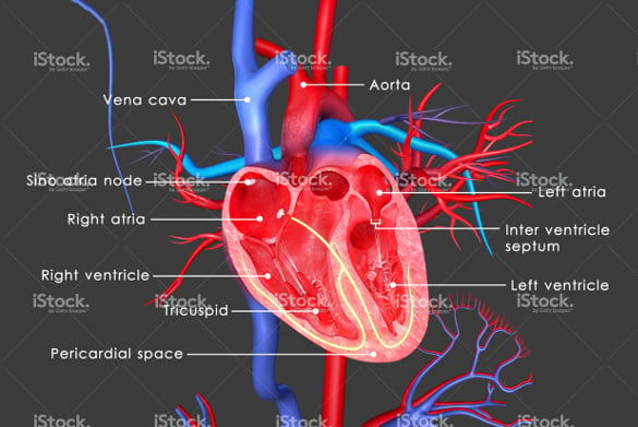
Diagram of Human Heart and Blood Circulation in It The outermost layer of your heart wall is called the epicardium, which is basically a very thin layer of serous membrane. The membrane provides lubrication and protection to the outer side of your heart, as you can see in heart diagram labeled. Myocardium Right beneath epicardium is another relatively thicker layer called myocardium.
Cat Anatomy and Physiology 101: All You Need to Know A - cervical bones, B - thoracic bones, C - lumbar bones, D - sacral bones, E - tail bones, 1 - cranium, 2 - mandible, 3 - scapula, 4 - sternum, 5 - humerus, 6 - radius, 7 - phalangeals, 8 - metacarpals, 9 - carpal bones , 10 - ulna, 11 - ribs, 12 - patella, 13 - tibia, 14 - metatarsals, 15 - tarsal bones, 16 - fibula, 17 - femur
How the Heart Works - The Heart | NHLBI, NIH - National Institutes of ... The Heart. The heart is an organ about the size of your fist that pumps blood through your body. It is made up of multiple layers of tissue. Your heart is at the center of your circulatory system. This system is a network of blood vessels, such as arteries, veins, and capillaries, that carries blood to and from all areas of your body.
Frog Anatomy & Physiology: Learn About All Parts Of The Frog The circulatory system of the frog consists of a three-chambered heart, blood, blood vessels, and the spleen. The frog's heart has two upper chambers (atria) and one lower chamber known as the ventricle. The right atrium receives oxygen-poor blood from the body, and the left receives oxygen-laden blood from the lungs.



:max_bytes(150000):strip_icc()/heart_interior-570555cf3df78c7d9e908901.jpg)









(230).jpg)




Post a Comment for "41 external structure of the heart with labels"